Cardiogenic Field
In the middle of the third week, the cardiac progenitor cells and blood islands lie in the splanchnic layer of the lateral plate mesoderm. The underlying pharyngeal endoderm induces the cells to form the cardiac myoblasts (angiogenetic clusters). The blood islands unite to form a horseshoe-shaped plexus of blood vessels surrounded by the myoblasts. The ventral part of horseshoe plexus forms the cardiogenic region, and the later portions form the dorsal aortae. The embryonic cavity over the cardiogenic region develops later into the pericardia cavity.
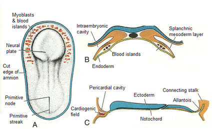
Human embryo - day 18: A. dosal veiw of embryo shows myoblasts and blood islands reside in the splanchnic mesoderm in front of the neural plate and on each side of the embryo. B. Transverse section show the position of the blood islands in the splanchnic mesoderm layer. C. Cephalocaudal section showing the position of the pericaridal cavity and cardiogenic field (Sadler 2010).
Position
Initially, the cardiogenic region lies in front of the oropharyngeal membrane and neural fold. Brain growth and cephalic folding of the embryo pulls the oropharyngeal membrane forward. As a result, the heart and the pericardial cavity move to the thorax.
Initially, the cardiogenic region lies in front of the oropharyngeal membrane and neural fold. Brain growth and cephalic folding of the embryo pulls the oropharyngeal membrane forward. As a result, the heart and the pericardial cavity move to the thorax.
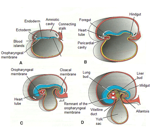
figures showing effects of the rapid growth of the brain on positioning of the heart. Initially, the cardiogenic area and the pericardial cavity in front of the oropharyngeal membrane. A. 18 days. B. 20 days. C. 21 days. D. 22 days. (Sadler 2010)
