Formation of The Heart Tube
Cardiogenic clusters in splanchic mesoderm are canalized to form paired heart tubes. As a result of embryo cefalocadual and lateral folds, the heart tubes fuse and form single heart tube. The heart tube remains attached dorsally to the pericardial cavity by the dorsal mesocardium that disappear with time and forms transverse pericardial sinus. So, the blood vessels suspend the heart tube in the pericardial cavity.
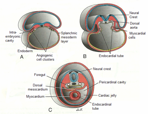
Transverse section through embryos at different stages of development, showing formation of a single heart tube from paired primordia. A. 17 days old embryo. B.18 days old embryo. C. 22 days old embryo. Fusion occurs only in the cadual region of the horseshoe-shaped tube. The crescent portion of the hoseshoe forms the outflow tract and most of the ventricular region (Sadler 2010)
Heart Layers
All the heart layers are derived from the splanchic mesoderm around the heart tube, except part of the epicardium which derived from mesothelial cells on the surface of the septum transversum. The endocardium lines the heart tube, the myocardium forms the muscular wall in the middle, and epicardium or visceral pericardium covers the outside of the heart tube.
